Monday 1 October 2012
DNA and Social Responsibility: Raymond Gosling: Flickr set dedicated to PhD thesi...
DNA and Social Responsibility: Raymond Gosling: Flickr set dedicated to PhD thesi...: Following the successful digitisation of Raymond Gosling's PhD thesis, 'X-ray diffraction studies of Deoxyribose Nucleic Acid' I have add...
DNA and Social Responsibility: Raymond Gosling: Visit to King's Archives
DNA and Social Responsibility: Raymond Gosling: Visit to King's Archives: A few months ago, I had the pleasure of meeting Professor Raymond Gosling as he came into the Archives to be interviewed for a Swedish do...
DNA and Social Responsibility: Raymond Gosling: Not just an extraordinary envoy
DNA and Social Responsibility: Raymond Gosling: Not just an extraordinary envoy: Raymond Gosling was one of the key workers on DNA at King's during the period that became immortalised as the 'Race for the Double Hel...
Friday 7 September 2012
DNA and Social Responsibility: Not another conference: A letter from Sir Nevill M...
DNA and Social Responsibility: Not another conference: A letter from Sir Nevill M...: While trawling through the papers relating to political activism it is difficult it is difficult to keep track of the myriad political gro...
Wednesday 5 September 2012
Digitising X-rays: the digitisation of the Biophysics collection
In this week’s post, I discuss some of the digitisation
aspects of the project with special reference to the work of one of our
digitisation contractors, MAX (previously MAX COMMUNCATIONS,) who digitised 4,000
of the glass and acetate material.
In October 2011, two of the digitisers from Max Ltd visited the archives in order to
familiarise themselves with the collection and carry out some test scanning.
The material was quite diverse: photographic prints, x-ray acetates, various
sizes of glass plate negatives, folded negative rolls and 35mm mounted slides.
The majority of this material required external specialists with the required
expertise and equipment to undertake the scanning. A small test file was
created composed of quarter plate glass negatives and x-ray acetates and sent
to the Wellcome for approval. Both Iain Stringer, who would go on to digitise
the collection, and David Cordery, head of Max
Ltd, have prior professional experience of working with glass plate and
acetate x-ray collections at various institutions around the UK and we were fully confident of
their ability to handle the fragile items and successfully scan them. The test
images that were sent to the Wellcome were approved and scanning commenced in
December 2011.
The images were
scanned at 300 dpi (dots per inch) at 8-bit (bit rate) RGB (Red Green Blue)
using an Epsom V750 Pro scanner. This type of flatbed scanner is reliable and
fast and from a preservation perspective, the scanner was suitable for digitising
the x-ray acetates as the two- inch gap between the bed of the scanner and the
top scanner head meant that it did not press onto the x-rays and so would not
cause any further damage to the x-rays afflicted with vinegar syndrome (vinegar
syndrome occurs when an acetate degrades and begins to oxidise creating a
vinegar smell. The surface often begins to warp and crack and this eventually
affects the emulsion. Unfortunately the process is irreversible and it is why
digitisation is one of the most effective ways of preserving an accessible copy
of the item.)
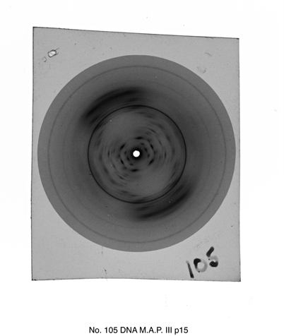 |
| An example of an X-ray acetate diffraction image from the collection. The sleeve caption information has been added to the image. This diffraction image was taken by Wilkins around 1953-1955 and the likely source of the DNA originated from human subjects supplied by Leonard Hamilton and Ralph Barclay of the Sloan Kettering Cancer Center in New York. |
I asked Iain to tell me about his experience scanning our
material compared to his previous experience with similar collections. He said
that the plates, in terms of general condition, were some of the best that he
has worked with as hardly any were chipped or broken. The only slight issue
that he encountered was that some of the slides were mounted with red strips,
the adhesive of which had begun to seep and caused them to attach themselves to
their transparent sleeves. In such cases, he therefore had to carefully remove
the slide from its sleeve. This required a degree of perseverance, depending on
the age and location of the adhesive strips on the slide.
Regarding the x-ray acetates, I had assumed that this
material would be trickier to scan considering the conditions that some of them
were in. Iain surprised me by saying that for the purposes of scanning they
were quicker to scan than the glass plates. Whilst care had to be taken in
handling small, fragile and brittle objects like deteriorating x-ray acetates,
the most time consuming element of scanning an x-ray was the post-production.
MAX Ltd provided us with images in three formats: the raw TIFF original file, the enhanced TIFF
amended file and a JPEG file. While enhancing an image can be difficult with
regard to obtaining an authentic copy of the original, in a situation where the
original is difficult to discern, post-production ‘clean up’ is necessary. The
majority of the x-ray acetates retained a degree of visible content and by
using Adobe Photoshop post production, it made it easier to enhance the original
pattern of the x-ray and compensate for some of the surface damage caused by
any deterioration.
Finally, I asked Iain what he thought of the acetate and
glass material as a whole. He said:
“ I found the material
quite interesting…I’ve learnt more about DNA than I have since school,, good
thing about my job that I don’t have to concentrate on one specific thing.
X-rays of DNA, diffraction, very interesting. They would definitely make a good
print, stretched over a canvas, especially one of the really clear ones like
‘Photo 51’”
I agreed, diffraction patterns such as ‘Photo 51’ are
visually striking though I personally am more in awe about the crystalline
A-form DNA pictures as there is something rather mesmerising about the symmetry
of these patterns. You can judge for yourself however, as these two x-ray
patterns are shown below.
 |
| A-form DNA |
 |
| B-form DNA |
Sunday 19 August 2012
Will the archives of the future be made of the strand of DNA?
The prospect of combining archives and DNA feels like a plotline of a Twilight Zone episode. What's exciting however is that it is a distinct possibility. The Guardian have just released a story about the DNA inscription of a book ( http://www.guardian.co.uk/science/2012/aug/16/book-written-dna-code) initially reported in the US journal, Science. The book composed of 53,000 words includes eleven images and a computer program. The 5.27 megabit collection of data created over several days was produced by Professor George Church of Harvard Medical School.
The method they used, in principle, was the same as digital inscription: encoding all the book information into a binary sequence. The DNA base pairs in this case representing 1's and 0's with Arginine (‘A’) and Cytosine (‘C’) representing zero, and Guanine (‘G’) and Tyrosine (‘T’) representing one. The team developed a system in which an inkjet printer embeds short fragments of artificially synthesized DNA onto a glass chip. Each DNA fragment contains a digital address code that denotes location within the original fiDNAle.
What makes DNA such a brilliant medium for storage is its data storage with estimates suggesting a gram of DNA can store 455 billion gigabytes. The data is easily readable and copied and maintains its stability for several thousand years.
The possibilities are fantastic. To put this in perspective, most digital formats require an upgrade after five years with physical data storage such as DVDs having, at most, a twenty year life span. This is because of the constant change in informational software packages within sturdy digital formats, such as TIFFs and PDFs having 10-15 year maximum life span. A DNA code sequence is therefore more desirable than a digital approximate, but neverless it is an exciting development and will potentially rival the paper record revolution in record keeping. This is an exciting archival perspective.
As a cataloguer and digitiser of the DNA related material of the Kings college London archive such a development is one of a personal joy. It would feel wonderfully apt to have the papers charting the discovery of the structure of DNA are encoded into DNA for future generations.
Tuesday 7 August 2012
August Project Update
The project is entering its final months and a quick update as it what has been happening is in order:
To date, 24,000 images have been produced by our digitisers which roughly breaks down as 4000 glass plate and acetate images and 20,000 images from the paper collection. Over the next two months the remaining part of the paper collection will be scanned. Sections that have already been scanned include papers from Wilkins’ early life, scientific working papers, correspondence with scientific colleagues, papers associated with the history of the research on DNA and sections of his autobiography.


Above, is a low resolution copy of some of the images that are being produced. The example is a postcard received by Maurice Wilkins from Francis Crick dated May 1955 and sent from Paris. The postcard reads: "Having a lovely time telling people about your work and my ideas! Hoping to see you in Cambridge for a quiet weekend - Francis".
Our main tasks over the next few months involve the construction of metadata and copyright and sensitivity checking. The latter is the most time consuming as a detailed survey requires a systematic check of all potentially risky material. Our catalogue descriptions are written at a level to summarize the contents of the physical file but because the images will be accessible individually an item level approach to sensitivity and copyright is needed to be certain that legally and ethically all necessary precautions are taken before publishing on-line. Needless to say this process is time-consuming and has proved to be the most taxing element of the project.
Apart from the construction of metadata and the sensitivity checking the only other main strand of the project to update everyone with is outreach. The project continues to gather interest from its social media sites (like the one I’m writing on now). Besides the blogs, the project has had a presence on Twitter and new images have been added to the project’s Flickr site. In May, the archives participated in a Radio 4 piece on the Wellcome Digital Library which was reported previously on this blog. Alongside this, we were privileged to have been visited by Raymond Gosling in March as part of a television documentary. It was wonderful to meet a contemporary of Wilkins and Franklin and hear from one of the key workers his own experience working at King’s at the time. We are quite fortunate to have a copy of Gosling's original 1954 PhD thesis, titled ''X-ray diffraction studies of Deoxyribose Nucleic Acid' which has been selected to be digitised as part of the King's College London Biophysics collection.
Subscribe to:
Posts (Atom)