In this week’s post, I discuss some of the digitisation
aspects of the project with special reference to the work of one of our
digitisation contractors, MAX (previously MAX COMMUNCATIONS,) who digitised 4,000
of the glass and acetate material.
In October 2011, two of the digitisers from Max Ltd visited the archives in order to
familiarise themselves with the collection and carry out some test scanning.
The material was quite diverse: photographic prints, x-ray acetates, various
sizes of glass plate negatives, folded negative rolls and 35mm mounted slides.
The majority of this material required external specialists with the required
expertise and equipment to undertake the scanning. A small test file was
created composed of quarter plate glass negatives and x-ray acetates and sent
to the Wellcome for approval. Both Iain Stringer, who would go on to digitise
the collection, and David Cordery, head of Max
Ltd, have prior professional experience of working with glass plate and
acetate x-ray collections at various institutions around the UK and we were fully confident of
their ability to handle the fragile items and successfully scan them. The test
images that were sent to the Wellcome were approved and scanning commenced in
December 2011.
The images were
scanned at 300 dpi (dots per inch) at 8-bit (bit rate) RGB (Red Green Blue)
using an Epsom V750 Pro scanner. This type of flatbed scanner is reliable and
fast and from a preservation perspective, the scanner was suitable for digitising
the x-ray acetates as the two- inch gap between the bed of the scanner and the
top scanner head meant that it did not press onto the x-rays and so would not
cause any further damage to the x-rays afflicted with vinegar syndrome (vinegar
syndrome occurs when an acetate degrades and begins to oxidise creating a
vinegar smell. The surface often begins to warp and crack and this eventually
affects the emulsion. Unfortunately the process is irreversible and it is why
digitisation is one of the most effective ways of preserving an accessible copy
of the item.)
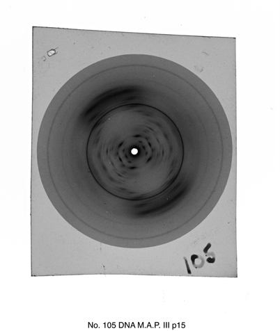 |
| An example of an X-ray acetate diffraction image from the collection. The sleeve caption information has been added to the image. This diffraction image was taken by Wilkins around 1953-1955 and the likely source of the DNA originated from human subjects supplied by Leonard Hamilton and Ralph Barclay of the Sloan Kettering Cancer Center in New York. |
I asked Iain to tell me about his experience scanning our
material compared to his previous experience with similar collections. He said
that the plates, in terms of general condition, were some of the best that he
has worked with as hardly any were chipped or broken. The only slight issue
that he encountered was that some of the slides were mounted with red strips,
the adhesive of which had begun to seep and caused them to attach themselves to
their transparent sleeves. In such cases, he therefore had to carefully remove
the slide from its sleeve. This required a degree of perseverance, depending on
the age and location of the adhesive strips on the slide.
Regarding the x-ray acetates, I had assumed that this
material would be trickier to scan considering the conditions that some of them
were in. Iain surprised me by saying that for the purposes of scanning they
were quicker to scan than the glass plates. Whilst care had to be taken in
handling small, fragile and brittle objects like deteriorating x-ray acetates,
the most time consuming element of scanning an x-ray was the post-production.
MAX Ltd provided us with images in three formats: the raw TIFF original file, the enhanced TIFF
amended file and a JPEG file. While enhancing an image can be difficult with
regard to obtaining an authentic copy of the original, in a situation where the
original is difficult to discern, post-production ‘clean up’ is necessary. The
majority of the x-ray acetates retained a degree of visible content and by
using Adobe Photoshop post production, it made it easier to enhance the original
pattern of the x-ray and compensate for some of the surface damage caused by
any deterioration.
Finally, I asked Iain what he thought of the acetate and
glass material as a whole. He said:
“ I found the material
quite interesting…I’ve learnt more about DNA than I have since school,, good
thing about my job that I don’t have to concentrate on one specific thing.
X-rays of DNA, diffraction, very interesting. They would definitely make a good
print, stretched over a canvas, especially one of the really clear ones like
‘Photo 51’”
I agreed, diffraction patterns such as ‘Photo 51’ are
visually striking though I personally am more in awe about the crystalline
A-form DNA pictures as there is something rather mesmerising about the symmetry
of these patterns. You can judge for yourself however, as these two x-ray
patterns are shown below.
 |
| A-form DNA |
 |
| B-form DNA |
No comments:
Post a Comment